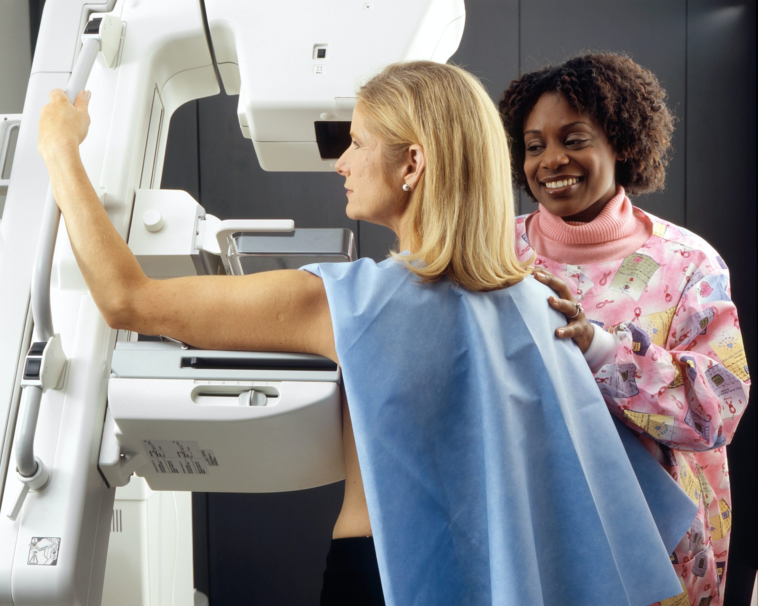
A mammography This is important evidence for diagnosis Breast cancerConsequently, they are performed not only in patients who have a suspicion of a tumor. They are also very useful in annual screening to detect cancer as soon as possible. Nevertheless, many women are afraid of this test from the pain that she usually causes. The chest is introduced between the two plates and pressed so that the X -Rays can go through them correctly. When pressing them, There may be a lot of pain The test is quite annoying. All this, without saying that, even being the minimum amount of X -RAYS, its frequent use can become dangerous. For all this, the operation Ultrasound computerized tomographyA test in the dough phase that promises to solve both problems.
This method is the axis on which the European QUSTOM project is coordinated Barcelona supercomputer centerFrom the National Supercomputer Center. Generally speaking, it consists of a device that does not use X -RAYS, but UltrasoundSo this is completely safe. In addition, its method of use is very convenient and does not imply any pain. Evidence of confirmation of its effectiveness began in 2024, in Vall d’Hebron HospitalIt is not yet available for generalized use by the field, since it is important to make sure that this is a really good alternative to mammography. However, with these first tests it is demonstrated that this can Even to be more accurate.
And, in fact, mammography also has an additional handicap for patients with Very fibrous or compact breasts. In these cases, X -rays can show the inside of the chest as a kind of white place. Since you can also see tumors, it is quite possible that breast cancer Incorrectly. This does not seem to happen with ultrasonic computerized tomography. Therefore, this may be the second cause of unsurpassed for mamographers in the future. If the tests are still positive, of course.
How does ordinary mammography work?
To conduct mammography, the chest is located between two plates and pressed. Then go through A small amount of X -RayThis is to go to the detector who composes with them images that can be printed as it is or send to the computer where they are digitized, and the group to cause a more complete image of the inside of the chest.
What is the difference with ultrasonic computed tomography?
Ultrasound computerized tomography analyzes the inside of the chest much less invasive. The patient should lie on a stretcher in which there are two deposits with water at 36.5 ºCAs soon as the chest is introduced in these deposits, ultrasounds are launched; That is, high -frequency sound waves that pass through fabrics or structures that want to analyze. In this case, the chest.
An analysis of the reflection of these waves when passing through the fabric can give an idea of their condition, as well as possible anomalies. Usually a Driver gel So the ultrasound can go to the area to be visualized. For example, a typical gel that is placed in the stomach for pregnant women. However, in this case, a moderate water bath, in which the chest is introduced by the driver’s gel.
Enough with 3 minutes for each chest To get a quick, safe and painless result.
Problems of three -dimensionality
Another of the great strengths of ultrasound computerized tomography is the use of supercomputing for obtaining More accurate images. Through ultrasound, many images have been received that go to a computer that reconstructs them to lead to 3D image Much more precise.
In 2024, he began to take place in Vall d’Hebron A Calculation of validationThe field, that is, the calculation of the forecast value of the test for a specific clinical result. In fact, in this case, its ability to detect breast cancer is measured.
It seems that this is quite effective, but additional research is still needed. At the moment, ordinary mammography remains necessary; But, I hope, in the future, these necessary evidence can be made without suffering. The time has come.
Source: Hiper Textual


