


In a study published in the journal Communications Biology, EMPA, researchers from Switzerland and St. Gallen, Explain how atomic force microscopy can be an ally for assessing Alzheimer’s disease progression.
Always in the race for early detection, researchers are working to obtain more effective techniques for diagnosing and treating neurodegenerative diseases.
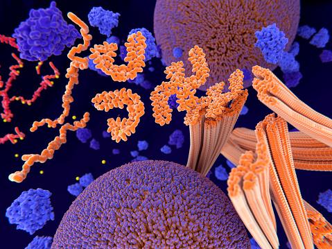
Alzheimer’s disease is the most common type of dementia and mainly affects people over the age of 65. It is formed by the accumulation of Tau and beta-amyloid proteins in neuronal cells.
Learn more: How is Alzheimer’s treated?
Trigger factors for the abnormal accumulation of these proteins are still under investigation. There is no cure for the disease. However, it is known that the earlier the patient receives the diagnosis, the higher the chance of managing and controlling the disease.
How is disease progression analyzed?
A still unanswered question was how to analyze how far the disease had progressed.
In clinical and laboratory tests, cognitive losses or the amount of proteins in the blood or cerebrospinal fluid can be analyzed, but not how far the disease has progressed. But EMPA researchers may have found another ally for that purpose.

Researchers who analyzed the size of protein fibrils found in the cerebrospinal fluid (CSF) of 33 patients with the disease may have found something promising.
Using atomic force microscopy, they were able to analyze the composition and size of the fibrils at the nanoscale.
By comparing the material collected from the participants, they were able to observe that these components do not come together in the CSF of healthy people, or that the protein fibrils are short and simple.
However, the size and complexity of the fibrils was remarkable in Alzheimer’s patients.
The researchers observed that patients with more signs and symptoms had larger and entangled fibers, thus linking disease progression to the complexity found in fibrillar structures.
For scientists, the new technique could be another tool for analyzing and diagnosing Alzheimer’s disease, even in its early stages, through analysis of the composition of the fibrils.
The team plans to use this technique to analyze fibrils in blood samples to increase the number of participants in subsequent tests and to further facilitate diagnosis.
Source: Tec Mundo
I’m Blaine Morgan, an experienced journalist and writer with over 8 years of experience in the tech industry. My expertise lies in writing about technology news and trends, covering everything from cutting-edge gadgets to emerging software developments. I’ve written for several leading publications including Gadget Onus where I am an author.


: Everything we know a week after its release")


