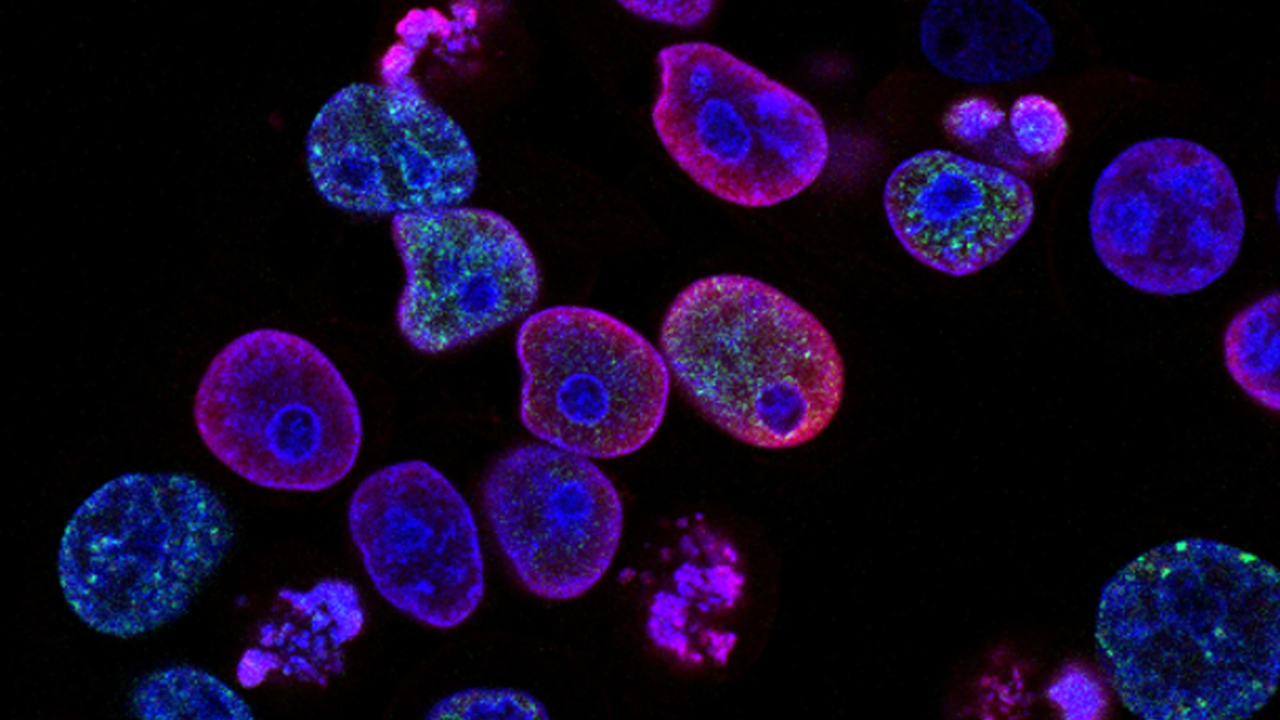
The material was filmed by them using the latest real-time visualization methods for this. As part of their latest study, the researchers looked inside the cells of a developing fruit fly.
To do this, they needed the “help” of ultra-high-resolution fluorescent microscopy techniques. With its help, scientists filmed how a cell grows, with a fine network of protein fibers under its surface, each 10,000 times thinner than a human hair.
It is reported that such observations may help to better understand wound healing processes in humans and how abnormalities develop in children with birth defects.
Source: Ferra


